Shamira Shahar, MB BCH BAO (Ireland)
Medical Officer, Diabetes and Endocrine Unit
Department of Medicine, Hospital Putrajaya,
Jalan P9, Presint 7, 62250 Putrajaya, Malaysia
Tel. No.: +60129380300
E-mail: shamira.shahar@gmail.com
ORCiD: https://orcid.org/0000-0003-1797-5237
e-ISSN 2308-118x
Printed in the Philippines
Copyright © 2019 by the JAFES
Received August 7, 2019. Accepted October 11, 2019.
Published Online First: November 9, 2019.
Eight cases of parathyroid carcinoma were identified (8 females; median age 45 years, range 28-72). Half of whom were diagnosed preoperatively. Hypercalcemic symptoms were seen in 87.5% of the patients and the main complication was nephrolithiasis. At presentation, the median calcium was 3.675 mmol/L, median phosphate of 0.68 mmol/L, median intact parathyroid hormone (iPTH) was 211 pmol/L. Five patients had regional nodes metastasis and 1 had distant metastasis to the lungs. Parathyroid gland invasion to adjacent structures was seen in 62.5% of cases while another 62.5% showed capsular or vascular infiltration on histology with median tumour size of 3.2 cm. Recurrent hypercalcemia occurred in 50% of the patients with median time of recurrence of 21 months. In this case series, we found that patients with severe hypercalcemia and high iPTH also exhibited a high index suspicion of PC.
Keywords: parathyroid neoplasms, carcinoma, hypercalcemiaParathyroid carcinoma (PC) is a rare malignancy. It accounts for 1% of patients with primary hyperparathyroidism. It was first described in 1904.[1] 90% of PC is functional and sporadic. It can be a part of the genetic or familial syndrome including Multiple Endocrine Neoplasm (MEN)1, MEN2A, isolated familial hyperparathyroidism and hyperparathyroidism jaw syndrome. The presentation is usually due to complications of hypercalcemia such as bone disease, renal disease or hypercalcemic crisis.[2] Yet, PC remains a challenge due to its rarity and lack of distinct features as compared to benign primary hyperparathyroidism. This is a pilot study conducted in Malaysia to identify the clinicopathologic features of PC and its morbidity and survival in Hospital Putrajaya, one of the leading endocrine institutions in Malaysia.
We retrospectively reviewed patients diagnosed with PC in Hospital Putrajaya from 2002 to 2018. Data were collected from the electronic medical records by searching for patients who had an International Classification of Diseases, Tenth Revision code for PC. All patients selected in this case series were diagnosed either postoperatively by histology, or based on clinical presentation and radiological images. Data including demographic, clinical, biochemical including calcium and parathyroid hormone (PTH) levels, pathologic characteristics, treatments including surgery and radiation therapy, complications, recurrence, and mortality were collected.
There were eight patients identified in our review, with a median age of 45 years (range 28-72), all were female. Diagnosis of PC was made preoperatively in 50% of patients based on clinical suspicion. Neck swelling was seen in 37.5% and 87.5% had hypercalcemic symptoms including polyuria and polydipsia, abdominal pain, bone pain, and weight loss. All patients presented with hypercalcemia complications including osteoporosis, fractures, nephrolithiasis, pancreatitis, and brown tumours. Median calcium at presentation was 3.675 mmol/L (range: 3.05-4.04), serum phosphate of 0.68 mmol/L (range: 0.58- 1.22), serum iPTH 211 pmol /L (range: 72-366) (Table 1).
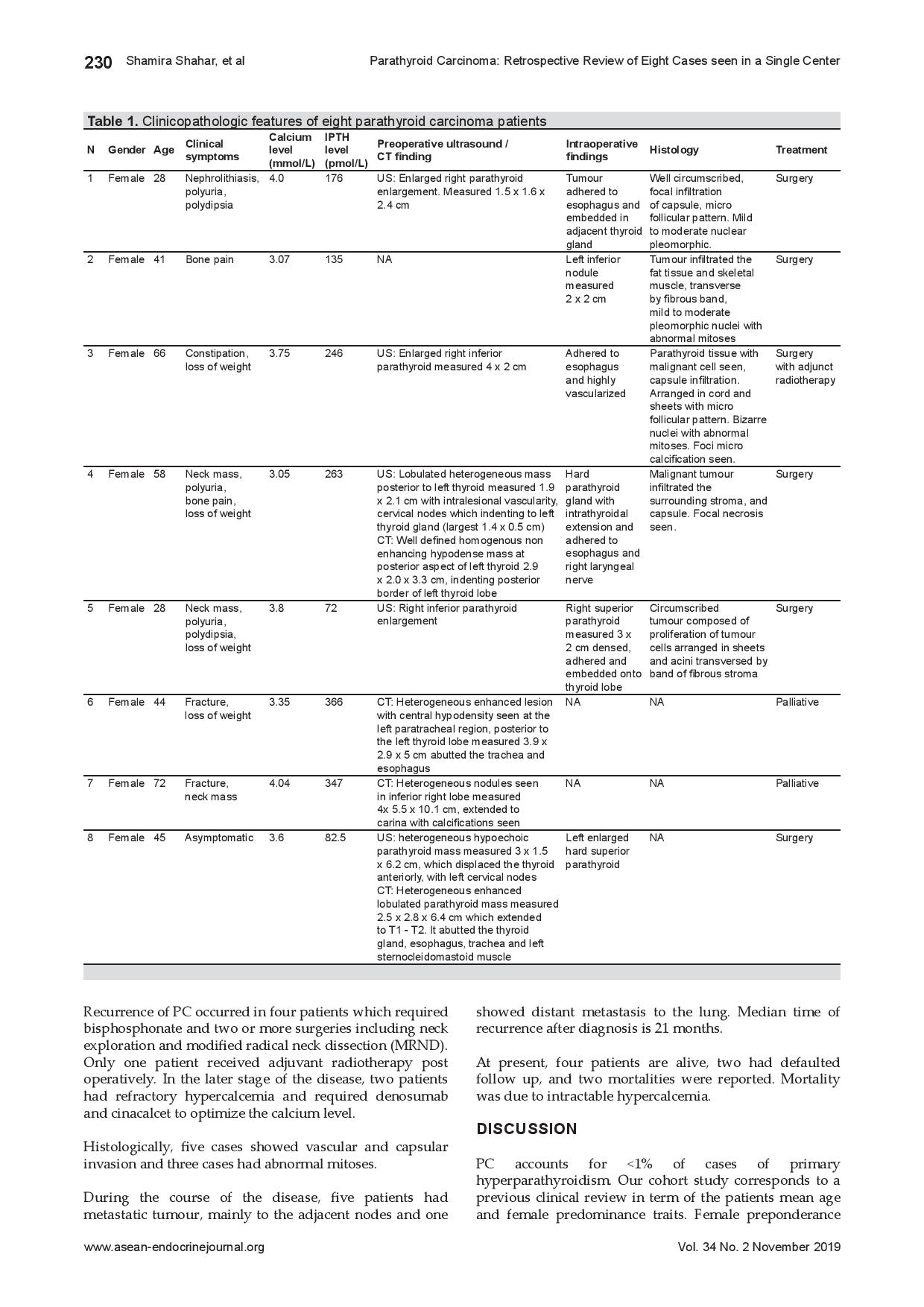
Table 1. Clinicopathologic features of eight parathyroid carcinoma patients
Ultrasound (US) is the most common imaging procedures used prior to operation. In our case series, 6 neck US was carried out. The analysis of the US findings revealed the characteristics of PC in two patients which includes heterogeneous hypoechoic lesion and presence of intralesional vascularity. In addition, 4 patients underwent CT of the neck and thorax preoperatively and demonstrated median tumour size of 3.2 cm, heterogeneous lesion, calcifications and tumour abutting the adjacent structures.
Surgery was performed in six patients. Two had en bloc resection and four had simple parathyroidectomy at first encounter. The two patients who had en bloc resection were diagnosed with PC preoperatively while others were not. Remaining two patients opted for medical therapy due to advanced age and inoperable tumour. All patients who underwent operation required bisphosphonate and one patient required additional calcitonin to optimize the calcium level. Intraoperatively, five cases show tumour invasion to its adjacent structures including thyroid lobe, esophagus, and recurrent laryngeal nerve. Four patients had developed hungry bone syndrome that required intravenous calcium gluconate postoperatively. One patient developed vocal cord paralysis after a second operation for recurrence, and 1 had surgical site infection.
Recurrence of PC occurred in four patients which required bisphosphonate and two or more surgeries including neck exploration and modified radical neck dissection (MRND). Only one patient received adjuvant radiotherapy post operatively. In the later stage of the disease, two patients had refractory hypercalcemia and required denosumab and cinacalcet to optimize the calcium level.
Histologically, five cases showed vascular and capsular invasion and three cases had abnormal mitoses.
During the course of the disease, five patients had metastatic tumour, mainly to the adjacent nodes and one showed distant metastasis to the lung. Median time of recurrence after diagnosis is 21 months.
At present, four patients are alive, two had defaulted follow up, and two mortalities were reported. Mortality was due to intractable hypercalcemia.
PC accounts for <1% of cases of primary hyperparathyroidism. Our cohort study corresponds to a previous clinical review in term of the patients mean age and female predominance traits. Female preponderance was also reported in other studies. However, it has been previously reported that there is no association of gender in PC. This disease affected women and men in a 1:1 ratio, as compared to that of primary hyperparathyroidism, where there is a marked female predominance, with a ratio of 3-4:1. In terms of geographic, race or income level, there has been no report to show any disparity.[3]-[4]
Majority of PC are hormonally functional, and patients often exhibit symptoms and complications of profound hypercalcemia at presentation due to elevated parathyroid hormone. Clinical presentation of PC commonly resulted in excessive PTH secretion by the functioning tumour, rather than invasion of the tumour mass into its surrounding structure. It is often associated with simultaneous manifestation of renal and skeletal failure during initial presentation.[5] In our study, all of our PC patients manifested hypercalcemic complications, mostly in bone and kidney. In a recent series, benign primary hyperparathyroidism reported renal involvement in less than 20% of the cases, whereby in PC, renal colic is a common presenting complaint, with 56% with nephrolithiasis and 84% with renal insufficiency were reported in one recent series.[6],[7],[8] Neck mass has been reported between 40-70% in several studies and it is an important clinical finding in PC as it is rarely seen in benign parathyroid disease.[9]
Serum calcium and parathyroid hormone were significantly elevated in our cohort study, consistent with previous studies which reported mean serum calcium >3.5 mmol/L[10] and markedly elevated PTH, usually 3-10 times above the upper limit associated with PC. Radiographic imaging was frequently needed prior to surgery for localization, however the procedure is less useful in evaluation of malignancy potential. Large tumours especially more than 2 cm, hypoechogenicity, heterogeneous, irregular borders, calcifications and local invasion were reported in several case series where malignancy should have been suspected.[10],[11],[12] In our study, only 2 US were reported preoperatively as PC, indicating invasion to adjacent trachea, cervical nodes involvement and intralesional vascularity.
The pathologic diagnosis of PC is challenging. Histopathologic criteria to diagnose PC include trabecular growth pattern, thick fibrous trabeculae, mitotic figures, and invasion of its capsule and surrounding vessels and lymph nodes.[13] However, these features are not exclusive to PC. In our study, 1 case was diagnosed later after she had undergone 3 surgeries for recurrent hypercalcemia. Therefore, it is crucial to note that the diagnosis of PC cannot depend primarily on histologic criteria as it shares the same features with benign parathyroid disease. Intraoperative findings are also essential to support diagnosis of PC. This includes lobulated firm mass, adhesion to thyroid lobe or adjacent cervical tissue such as strap muscles, recurrent laryngeal nerve, oesophagus and trachea.[14]
Genetic mutations such as CDC73 has been recognized to play an important role in the pathogenesis of PC. CDC73 is responsible to encode for parafibromin, which is involved in the regulation of gene expression and cell proliferation inhibition. It can be found in other mutated genes such as PIK3CA, MTOR, ADCK1, FAT3, AKAP9, and ZEB1, which were also identified in PC. Identification of these mutations carry important implications for management and early detection or prevention of PC among family members of PC patient.[15]
Surgery is the main treatment for PC. Complete surgical resection of tumour and involving tissue (en bloc) with microscopically negative surgical margins offers the best chance of cure and reducing the risk of recurrence. Studies have shown that preoperative suspicion and appropriate resection during primary surgery offers the best prognostic chance.[16],[17],[18] In the case of recurrence, surgical resection is still the primary mode of treatment. Significant palliation may result from the resection of recurrence lesions. However, repeated surgeries predispose patients up to a 60% lifetime accumulated surgical risk. To date, there is no established randomized trial available to evaluate radiotherapy and chemotherapy for PC. Incorporation of chemotherapy or radiotherapy needs to be tailored on individual basis.[19]
Hypocalcemia is a common immediate side effect of parathyroidectomy, despite being the cause of the disease. It can be severe, resulting in hungry bone syndrome, which is characterised by a rapid, profound and persistent hypocalcemia. Therefore, it is crucial to ensure that appropriate replacement of calcium and vitamin D are monitored closely. Hungry bone syndrome has been associated with advanced age, preoperative high level of serum calcium, ALP and iPTH, depleted vitamin D level and tumour size. In a randomized control trial among PHPT patients who were going for parathyroidectomy, daily supplementation of high dose vitamin D improves vitamin D status and decreases iPTH level preoperatively. However, this study found no association between preoperative 25(OH)D and postoperative serum Ca level with hungry bone syndrome. Even so, high dose of vitamin D supplement preoperatively is safe and improves bone mineral density and reduces bone resorption. Hence, in our center it is recommended for preoperative administration of vitamin D to minimize the need for prolonged intravenous calcium administration postoperatively.[20]
PC is a slowly progressing disease. It has a high recurrence rate of up to 49-60% of cases after the initial operation.[19] In our study, recurrence of the disease occurred in 4 patients, with mean time to recurrence was 21 months, while other studies reported 24-48 months. Metastasis usually occurs locally in 25-80%, and approximately 25% developed distant metastasis during the duration of the disease, most commonly in the neck, followed by the lung and spine.[21]
Mortality and morbidity in PC are mainly caused by hypercalcemia. Therefore, in treating PC, management needs to be focused on ameliorating the hypercalcemia effects. Apart from surgery, which has been the primary initial therapy in recurrent or refractory cases, medical therapy such as biphosphanate, calcimimetic, denosumab has also shown to control hypercalcemia. In a recent case report, denosumab has demonstrated a rapid normalization of calcium level in a case of refractory hypercalcemia secondary to recurrent PC.[22] Calcimimetic agent is approved by U.S. Food and Drug Administration (FDA) as the treatment for hypercalcemia in PC. One multi-centre study has demonstrated that calcimimetic has a durable clinical effect in achieving control of serum PTH and calcium levels in inoperable patients.[23]
Prognosis of PC is variable. Overall survival from various cancer databases showed of 85% and 49-77% at 5 and 10 years’ follow-up, respectively.[16] In several observational studies, mortality has been associated with metastasis, severity of hypercalcemia and lymph nodes involvement. Young age is associated with improved survival.[24],[25] Interestingly, a study of PC in a single institution revealed that survival rate and surgical complications were significantly reduced if initial operation was done in a dedicated endocrine centre versus other tertiary or nontertiary centres. Other studies reported early identification of the tumour and appropriate resection of the neoplasm at the time of primary surgery offered the patients the best prognostic chance.[12]
PC is a rare malignancy and has an indolent but progressive course. It remains a challenge to diagnose PC as it mimics benign primary hyperparathyroidism. Clinical judgement is crucial for early diagnosis of PC and to detect its recurrence and/or metastasis. Severe hypercalcemia (>3.5 mmol/L), very high iPTH (3-10 times upper limit) and large parathyroid lesion (>3 cm) should prompt high index suspicion of PC. Complete surgical resection with microscopically negative margins that can be reached with the en bloc excision is the best chance for cure. However, recurrence is not uncommon among PC survivor. Recurrence or metastatic disease should be surgically treated, and often multiple surgical interventions are needed, even though they are not definitely curative. Since PC is an indolent tumour with a long-lasting survival and the cause of death is mainly due to untreatable hypercalcemia, therefore, the main goal of therapy is to control hypercalcemia and its complications. Thus, lifetime follow up is mandatory.
Ethical ConsiderationAll information in the case series has been provided without mention of any identifier in an effort to ensure anonymity of the patients. The authors have sought ethical clearance from the Medical and Research Committee (MREC), Ministry of Health Malaysia (MOH) to conduct the study and publish the case series.
Statement of AuthorshipAll authors certified fulfillment of ICMJE authorship criteria.
Author DisclosureThe authors declared no conflict of interest
Funding SourceNone.
[1] De Quervain F. Parastruma maligna aberrata. Deutsche Zeitschr
Chir. 1904;100:334-52.
[2] Obara T, Fujimoto Y. Diagnosis and treatment of patients with
parathyroid carcinoma: An update and review. World J Surg.
1991;15(6):738-44.
[3] Shane E. Clinical review 122: Parathyroid carcinoma. J Clin Endocrinol
Metab. 2001;86(2):485-93.
[4] Favia G, et al. La patologia chirurgica della tiroide e delle
paratiroidi. Club delle UEC; Il carcinoma delle paratiroidi; 2000.
[6] Heath H 3rd, Hodgson SF, Kennedy MA. Primary hyperparathyroidism:
Incidence, morbidity, and potential economic impact in a community.
N Engl J Med. 1980;302(4):189-93.
[7] Silverberg SJ, Shane E, Jacobs TP, et al. Nephrolithiasis and bone
involvement in primary hyperparathyroidism. Am J Med. 1990;89(3):327-34.
[8] Wynne AG, van Heerden J, Carney JA, Fitzpatrick LA. Parathyroid
carcinoma: Clinical and pathological features in 43 patients. Medicine. 1992;71(4):197-205.
[9] Levin KE, Galante M, Clark OH. Parathyroid carcinoma versus
parathyroid adenoma in patients with profound hypercalcemia.
Surgery. 1987;101(6):649-60.
[10] Sidhu PS, Talat N, Patel P, Mulholland NJ, Schulte KM. Ultrasound
features of malignancy in the preoperative diagnosis of parathyroid
cancer: A retrospective analysis of parathyroid cancer. Eur Radiol.
2011;21(9):1865-73.
[11] Daly BD, Coffey SL, Behan M. Ultrasonography appearances of
parathyroid carcinoma. Br J Radiol. 1989; 62(743):1017-9.
Schantz A, Castleman B. Parathyroid carcinoma. A study of 70 cases.
Cancer. 1973;31(3):600-5.
[13] Delellis RA. Challenging lesions in the differential diagnosis of
endocrine tumors: Parathyroid carcinoma. Endocr Pathol. 2008;
19(4):221-5.
[14] Koea JB, Shaw JH. Parathyroid cancer: Biology and management. Surg
Oncol. 1999;8(3):155-65.
[15] Pandya C, Uzilov AV, Bellizzi J, et al. Genomic profiling reveals
mutational landscape in parathyroid carcinomas. JCI Insight.
2017;2(6):e92061.
[16] Sandelin K, Auer G, Bondeson L, Grimelius L, Farnebo LO. Prognostic
factors in parathyroid cancer: A review of 95 cases. World J. Surg.
1999;16(4):724-31.
[17] Schulte KM, Talat N, Galata G, et al. Oncologic resection achieving
r0 margins improves disease-free survival in parathyroid cancer.
Ann Surg Oncol. 2014;21(6):1891-7.
[18] Kebebew E, Arici C, Duh QY, Clark OH. Localization and reoperation
results for persistent and recurrent parathyroid carcinoma. Arch
Surg. 2001;136(8):878-85.
[19] Wei CH, Harari A. Parathyroid carcinoma: Update and guidelines
for management. Curr Treat Options Oncol. 2012;13(1):11-23.
[20] Rolighed, L, Rejnmark L, Sikjaer T, et al. Vitamin D treatment in
primary hyperparathyroidism: A randomized placebo controlled trial.
J Clin Endocrinol Metab. 2014;99(3):1072-80.
[21] Dudney WC, Bodenner D, Stack Jr BC. Parathyroid carcinoma.
Otolaryngol Clin North Am. 2010;43(2):441-53.
[22] Tong CV, Hussein Z, Noor NM, Mohamad M, Ng WF. Use of denosumab
in parathyroid carcinoma with refractory hypercalcemia, QJM.
2015;108(1):49–50.
[23] Silverberg SJ, Rubin MR, Faiman C, et al. Cinacalcet hydrochloride
reduces the serum calcium concentration in inoperable parathyroid
carcinoma. J Clin Endocrinol Metab. 2007;92(10):3803-8.
[24] Harari A, Waring A, Fernandez-Ranvier G, et al. Parathyroid
carcinoma: A 43-year outcome and survival analysis. J Clin Endocrinol
Metab. 2011;96(12):3679-86.
[25] Busaidy NL, Jimenez C, Habra MA, et al. Parathyroid carcinoma: A
22-year experience. Head Neck. 2004;26(8):716-26.
Authors are required to accomplish, sign and submit scanned copies of the JAFES Author Form consisting of: (1) Authorship Certification, that all the requirements for authorship have been met by each author, and that the final version of the manuscript has been read and approved by all authors; (2) the Author Declaration, that the article represents original material that is not being considered for publication or has not been published or accepted for publication elsewhere; (3) the Statement of Copyright Transfer [accepted manuscripts become the permanent property of the JAFES and are licensed with an Attribution-Share Alike-Non-Commercial Creative Commons License. Articles may be shared and adapted for non-commercial purposes as long as they are properly cited]; and the ICMJE form for Disclosure of Potential Conflicts of Interest. For original articles, authors are required to submit a scanned copy of the Ethics Review Approval of their research as well as registration in trial registries as appropriate. For manuscripts reporting data from studies involving animals, authors are required to submit a scanned copy of the Institutional Animal Care and Use Committee approval. For Case Reports or Series, and Images in Endocrinology, consent forms, are required for the publication of information about patients; otherwise, appropriate ethical clearance has been obtained from the institutional review board. Articles and any other material published in the JAFES represent the work of the author(s) and should not be construed to reflect the opinions of the Editors or the Publisher.